
Figure 1. Is the bias in attention in spatial neglect caused by an error in the gaze direction signals? Hypothesis in cake form, with thanks to the class 2018/2019 Psychology and Neuroscience at the University of St Andrews, YUM! & THANK YOU! ( Balslev and Odoj, Neuropsychologia, 2019 )
Figure 2. Does the sensory homunculus have two eyes? We used functional magnetic resonance imaging (fMRI) to map a proprioceptive projection from the eye muscles in the somatosensory cortex of both hemispheres. We found that the somatosensory homunculus on each side contains a representation of both eyes. This departs from the classic representation of the somatosensory homunculus where each side of the body is typically represented in the contralateral hemisphere only (Balslev, Albert and Miall, Human Brain Mapping, 2011; idea for figure suggested by Alex List)
Eye proprioception role in spatial attention
Our eyes relay a continuous stream of images to the brain. We are unaware of most of this input, because processing every detail would require resources that far exceed the available neural capacity. Attention is the mechanism by which the brain selects the most important features of the sensory streams and gives them a perceptual advantage.
A consistent observation in my experiments has been a change in the perception of visual stimuli when interfering with the activity of the eye proprioceptive area in the postcentral gyrus (Balslev, Gowen and Miall, J Cogn Neurosci, 2011, Odoj and Balslev, J Cogn Neurosci, 2013). The changes in perception are accompanied by changes in activity in the higher-order visual cortex (Balslev, Siebner, Paulson and Kassuba., Neuroimage, 2012). These effects are likely to be the result of the alteration of the oculoproprioceptive signal, because they can be replicated when the extraocular muscles are directly stretched (Balslev, Newman and Knox, Invest Opthalmology and Vis Science, 2012). My findings are intriguing because the somatosensory cortex is normally not associated with visuospatial attention. A role of this structure in attention however would explain a counter-intuitive observation. An extension of the lesion into the somatosensory cortex is associated with a milder attention bias in patients with spatial neglect (Balslev, Odoj and Karnath, J Neurosci, 2013).
How are the eyes and attention coupled?
One possibility is that the somatosensory cortex has a dual function, in coding eye position and in coding the locus of attention, like the oculomotor areas. Most oculomotor areas such as the frontal eye fields or the superior colliculi play a role in both moving the eyes and in moving the locus of attention.
Another possibility is that the somatosensory cortex contributes a proprioceptive gaze direction signal that is a component of the attention representations elsewhere. This signal aligns the retinotopic space to the physical space.
My research (Odoj and Balslev, J Cogn Neuroscience, 2016 ) supports the second hypothesis. After transcranial magnetic stimulation of the postcentral gyrus there is a consistent displacement of attention loci in the visual space relative to somatosensory cues. The shift is equal with the shift in perceived eye rotation and cannot be explained by a general mislocalization of the cues, because no errors are observed when participants are asked to reach to the visual targets, in the absence of visual hand feedback.
This research was funded by a Marie Curie fellowship for Career Development, a Research Grant from the Danish Medical Research Councils and an Institutional Strategic Support Fund at the University of St Andrews from the Wellcome Trust.
Could a perturbed gaze direction signal cause attention bias in unilateral spatial neglect?
Spatial neglect is a common and disabling condition in stroke survivors characterized by a lack of awareness of stimuli that appear in the contralesional (usually left) side of the space. A number of hypotheses have been proposed to explain disease mechanisms in spatial neglect, however, all these previous hypotheses would predict accurate perception in the right, ipsilesional hemispace. If spatial neglect is caused by an ipsilesional shift of the attentional priority map, however, one should be able to measure its signature throughout the visual field, including the "normal" hemispace. Our results confirmed this prediction. We found a small - one degree visual angle error between the locus of attention in the visual space and the somatosensory cue, the location of the patient's index finger, hidden from view. This is tiny, but it can be measured even in the normal hemispace.
This research was funded by a Research Grant from the Danish Medical Research Councils and an Institutional Strategic Support Fund at the University of St Andrews from the Wellcome Trust.
How can one study the role of the afferent signal from the eye muscles ?
It is very difficult to interfere with the sensory signal from the eye muscles (proprioception) in a controlled way. Until recently, the method used to dissociate the proprioceptive input from the oculomotor command in humans involved the application of a suction lens on the sclera to block the eye into a rotated position. Safety concerns obviously limit the use of this method. Following up on the identification of the eye proprioceptive area by invasive recordings in the monkey, my experiments in healthy humans showed that repetitive transcranial magnetic stimulation (rTMS) over the somatosensory cortex alters the perception of one's own eye position and reduces the ability to correct for a proprioceptive perturbation (Balslev and Miall, J Neurosci, 2008). The innovative use of rTMS, which provides a safe and painless method to study the role of the eye position signals in behavior and cognition, was acknowledged with the Young Investigator Award at the University of Oxford/Magstim Summer School in 2010 . This research was funded by a postdoctoral fellowship from the Danish Medical Research Councils.
Is it all about the efference copy or does eye proprioception matters too when estimating the direction of one's own gaze?
There are two main sources of eye position information. The efference copy of the command issued to the eye muscles or the corollary discharge and the afferent information from these muscles or eye proprioception. A role of the corollary discharge in visual localization has long been presumed because visual localization is intact after sectioning the nerves that brings proprioceptive input from the eye muscles to the brain. Proprioception was believed to play no role in visual localization, but rather update the corollary discharge over days. I had the opportunity to test this hypothesis in a patient with a rare, focal lesion of the somatosensory cortex. The results show that visual localization indeed relies on the corollary discharge. Proprioception however is continuously compared against the corollary discharge, to be incorporated into the eye position estimate as soon as a mismatch between the two signals is detected (Balslev and Miall, J Neurosci, 2008; Balslev et al., J Neurosci, 2012). This finding was the topic of a Young Investigator Talk at the Gordon conference of Eye Movement in 2011 and the paper describing it was awarded an Attempto Prize at the University of Tuebingen. This research was funded by a Marie Curie fellowship for Career Development and a Research Grant from the Danish Medical Research Councils.
A mismatch between prediction and re-afference occurs not only during growth or after surgery of the eye muscles in patients with strabismus, but also in everyday life, for instance every time one applies a contact lens. To dissociate the corollary discharge and eye proprioception in the laboratory, I use passive eye movement and repetitive transcranial magnetic stimulation (rTMS) over the eye proprioceptive area. In such conditions with an acute mismatch between the two eye position signals, the abnormal proprioceptive signal can be measured as an error in locating visual objects relative to the body
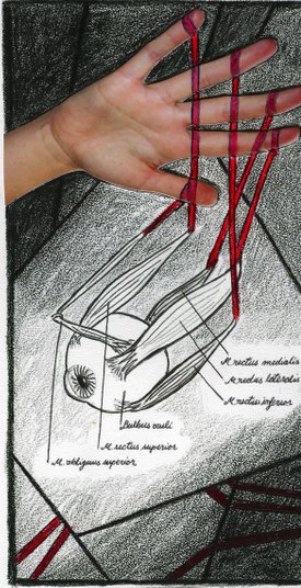
Figure 3. PhD thesis, a long time ago, (Hand) Proprioception - an obstacle for motor control in conditions with a visuoproprioceptive conflict, with thanks to Pauliina Aarnio for the cover illustration
Updated by Daniela on 16.06.2020 at 12:54